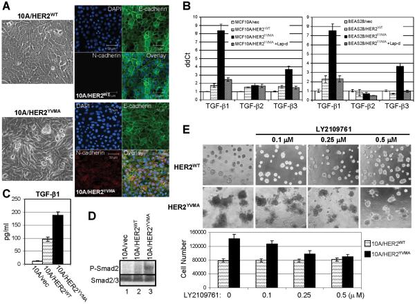
Fig. 1.
Autocrine TGF-β signaling is increased in cells expressing mutant HER2. A. Phase contrast (left) and immunofluorescence (right) images of MCF10A/HER2WT and MCF10A/HER2YVMA cells cultured to reach confluence. For immunofluorescence staining, cells growing on glass coverslips were fixed and stained using antibodies against E-cadherin (green) and N-cadherin (red). DAPI (blue), nuclear staining. Bar equals 50 μm. B. MCF10A (left) or BEAS2B (right) cells stably expressing HER2WT, HER2YVMA or empty vector were grown in complete medium and treated with lapatinib for 16 h or left untreated. Total RNA was extracted and subjected to reverse transcription followed by quantitative PCR for TGF-β1, -β2 and -β3 as described in Materials and Methods. Data were normalized to the MCF10A/vec or BEAS2B/vec control cells. Each data represents the mean ± S.D. of 3 experiments. C. Cells grown on 100-mm dishes (1×10⁶ cells/dish) were incubated for 24 h in serum-free medium. Conditioned medium (CM) was collected and analyzed for total amount of TGF-β1 by ELISA as indicated in Materials and Methods. Data are normalized to pg/ml/106 cells/24 h. Each data represents the mean ± S.D. of 3 experiments. D. Cells grown on 6-well plate were serum-starved for 16 h before lysed. Cell lysates were subjected to immunoblot using indicated antibodies. E. Top: MCF10A/HER2WT and MCF10A/HER2YVMA cells were plated in Matrigel in 8-well chambers and allowed to grow in the absence or presence of LY2109761 at the indicated concentrations. The inhibitor was added to the top medium 12 h after cell seeding and replenished every 3 days. Phase contrast images shown were recorded 9 days after the initial seeding of cells. Bottom: 9-day acini were trypsinized and total cell number was determined in a Coulter counter. Each bar graph represents the mean ± S.D. of 4 wells.

