Fig. 7.
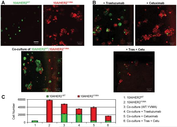
Combined inhibition of intracellular and paracrine effects of mutant HER2. A: MCF10A/HER2WT cells were labeled with PKH67 green fluorescent cell linker amd MCF10A/HER2YVMA cells were labeled with PKH26 red fluorescent cell linker. Labeled cells were immediately seeded in Matrigel for 3D culture in the absence of EGF in the top medium. Top: For single cell type culture, 6×10³ cells were seeded on day 0. Bottom: For co-culture of mixed cell types, 3×10³ cells of each cell type (a total of 6×10³ cells) were seeded on day 0. The fluorescent images were captured on day 6. Bars equal 100 μm. B: Co-culture of differently labeled 10A/HER2WT and 10A/HER2YVMA cells were set up as described in A. At 12 h after cell seeding, trastuzumab (10 μg/ml) and/or cetuximab (10 μg/ml) was added into the top medium as indicated. Antibodies were replenished every 3 days. The fluorescent images were photographed on day 6. C: The 6-day acini in A&B were trypsinized and total cell number of each labeled cell type was determined. Each bar graph represents the mean ± S.D. of 3 wells.

