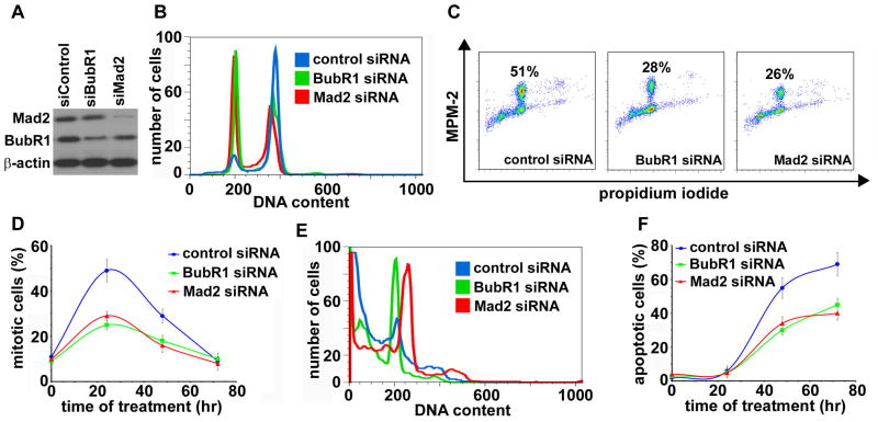
Figure 4.
EM011-induced apoptosis requires activation of spindle assembly checkpoint. Impairment of the spindle checkpoint by knock-down of Mad2 or BubR1 expression by specific siRNAs suppresses EM011-induced mitotic arrest and apoptosis. A. Immunoblot analysis of Mad2 and BubR1 expression in PC-3 cells transfected with luciferase (control), Mad2, or BubR1 siRNAs for 24 hr. Cells transfected with Mad2, BubR1, or control siRNAs for 24 hr were treated with 25 μM EM011 for 0, 24, 48, or 72 hr. B. Histogram-overlays of cell-cycle profiles of 24 hr drug-treated PC-3 cells that were transfected with luciferase (control), Mad2, or BubR1 siRNAs. C. Mitotic cells quantified by a mitosis-specific MPM-2 antibody in 24 hr drug-treated cells (transfected with luciferase (control), Mad2, or BubR1 siRNAs) using dual-color flow cytometry. D. Quantitation of MPM-2 positive cells in luciferase (control), BubR1 or Mad2 siRNAs transfected cells that were treated with EM011 for 0, 24, 48 and 72 hr. Values represent averages and error bars show SD (p < 0.05). E. Histogram-overlays of cell-cycle profiles of luciferase (control), BubR1 or Mad2 siRNA transfected cells that were drug-treated for 72 hr depicting differences in sub-G1 population (indicative of apoptotic cells). F. Quantitation of apoptotic cells (sub-G1) shows that cells with attenuated checkpoint molecules had reduced apoptotic responses at the noted hours (0, 24, 48 and 72) upon drug-treatment. Values represent averages and error bars show SD (p < 0.05).

