
Figure 4.
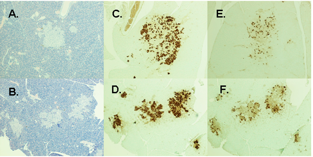
Representative images of pancreas sections from prediabetic sham and IT-operated animals at 2 months after surgery. Hematoxylin and eosin stain of pancreas sections from sham-operated (A) and IT-operated animals (B). Anti-insulin immunostaining of pancreas sections from sham-operated (C) and IT-operated (D) animals. Anti-glucagon immunostaining of pancreas sections from sham-operated (E) and IT-operated (F) animals.