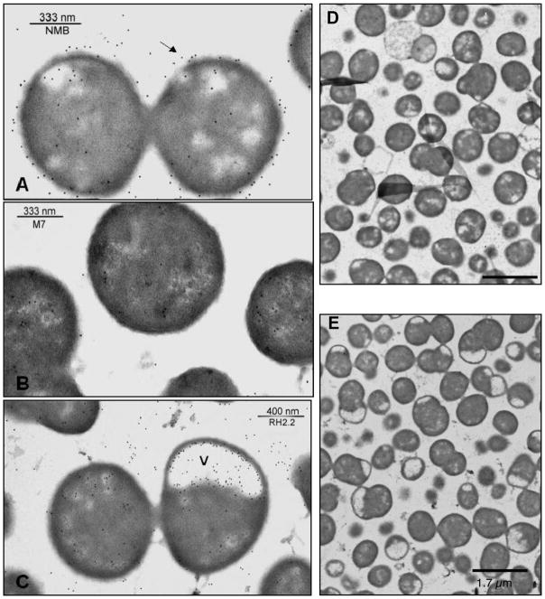
Fig. 4.
Electron photomicrographs of immunogold-labeled meningococcal cell sections. Panel (A) shows the wild-type parental strain NMB, (B) shows the unencapsulated mutant (synA) M7, and (C) shows the NMB0065 mutant. Labeling was performed with the serogroup B capsule-specific monoclonal antibody, 2-2-B, as the primary antibody, and the 10-nm gold-conjugated anti-immunoglobulin G/M antibody was used as the secondary antibody. The arrows indicate the capsule surrounding the wild-type strain but not the M7 or the NMB0065 mutant. The (v) indicates electron-translucent vacuoles containing internalized capsule of the NMB0065 mutant. Overviews of the wild type strain and the NMB0065 mutant are shown in Panels D and E, respectively.

