
Figure 1. Increased numbers of neutrophils, alveolar macrophages and T lymphocytes are present in alveoli of CF patients.
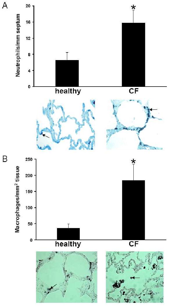
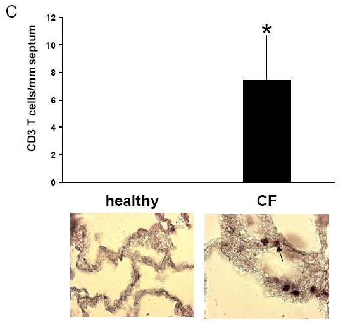
Neutrophils (A, arrow), stained with monoclonal antibodies directed against human neutrophil elastase, in CF alveolar lung tissue and in normal alveolar lung tissue differ significantly (p<0.0001) in numbers. Alveolar macrophages (B, arrow), stained with monoclonal antibodies directed against CD68 receptors, in CF alveolar lung tissue and in normal alveolar lung tissue differ significantly (p<0.0001) in numbers with regard to cells in septa (black bars) and intraluminally (white bars). T lymphocytes (C, arrow), stained with monoclonal antibodies directed against CD3, in CF alveolar lung tissue and in normal alveolar lung tissue differ significantly (p<0.0001) in numbers.