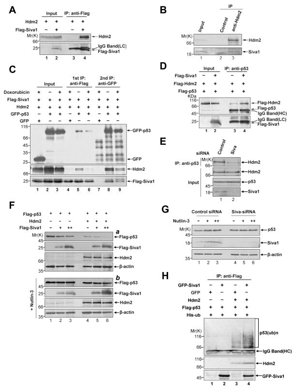
Figure 2. Siva1 binds to Hdm2 and enhances Hdm2-mediated p53 degradation.
(A) Association of ectopically-expressed Siva1 and Hdm2. p53−/−mdm2−/− MEF cells were transfected with Hdm2 and either Flag-Siva1 or vector control. After treatment with 20 μM MG132 for 6 h, whole cell lysates were prepared and immunoprecipitated with anti-Flag antibody. The lysates and immunoprecipitated proteins were analyzed by Western blotting.
(B) Association of endogenous Siva1 and Hdm2. Whole-cell extracts from U2OS cells treated with 20 μM MG132 for 6 h were immunoprecipitated with anti-Hdm2 or a control antibody (anti-HA), and followed by Western analysis using antibody against Hdm2 and Siva.
(C) Siva1, p53 and Hdm2 form a ternary complex. Flag-Siva1, Hdm2, GFP and GFP-p53 were transfected into p53−/−Mdm2−/− MEF cells in the indicated combinations. Cells were treated with 20 μM MG-132 for 2 h and with 4 μM doxorubicin or left untreated for an additional 6 h. Cell lysates were immunoprecipitated with EzviewTM Red anti-Flag M2 Affinity Gel (lanes 4–6). Flag-Siva1 and the associated proteins were eluted with 3XFlag peptide. Twenty percent of the eluent was subject to Western analysis using indicated antibodies. The remaining eluent was used for secondary immunoprecipitation with anti-GFP antibody (lanes 7–9).
(D) Overexpression of Siva1 enhances Hdm2-p53 interaction. H1299 cells were transfected with Flag-Hdm2, Flag-p53, and either Flag-Siva1 or the vector control. Transfected cells were treated with MG132 for 6 h. The association of Flag-p53 and Flag-Hdm2 was analyzed by immunoprecipitation assay with anti-p53 antibody.
(E) Knockdown of Siva1 diminishes the Hdm2-p53 interaction. The blot (top panel) shown in Figure 1I was re-probing with antibody against Mdm2. Input was equivalent to 10% of the whole cell lysates used for Co-IP.
(F) Siva1 affects p53 steady-state level in H1299 cells. H1299 cells were co-transfected with a fixed amount of Flag-p53 (0.1 μg), increasing amount of Flag-Siva1 (0, 0.1 and 0.3 μg), and together with either GFP-Hdm2 or GFP as indicated, then treated with (part b) or without (part a) Nutlin-3 (10 μM) for 24 h. Cell lysates were analyzed by Western blotting. Expression of β-actin was shown as a loading control.
(G) Siva1 affects p53 steady-state level via Hdm2. HCT116 cells were transfected with either Siva-siRNA or scramble-siRNA (control), and were then treated with increasing amount of Nutlin-3 (0, 5, 10 μM) for 24h. Cell lysates were analyzed by Western blotting. Expression of β-actin was shown as a loading control.
(H) Siva1 increases p53 polyubiquitination. p53−/−Mdm2−/− MEF cells were transfected with Flag-p53, His-ubiquitin, Hdm2, GFP-Siva1, GFP in the indicated combinations. Transfected cells were grown in medium containing MG132 (20 μM) for another 6 h. Flag-p53 was immunoprecipitated using anti-Flag antibody and analyzed by Western blotting using anti-p53 antibody.

