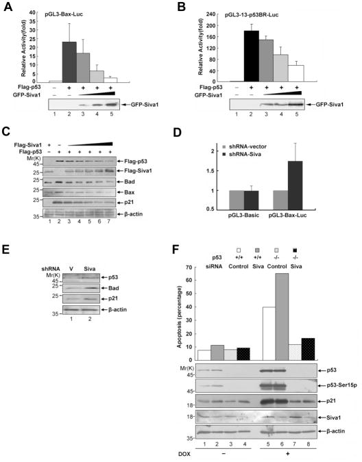
Figure 5. Siva1 suppresses p53-mediated gene expression and apoptosis.
(A) & (B) Overexpression of Siva1 inhibits the transcriptional activity of p53. H1299 cells were transfected with Flag-p53 (0.1 μg) together with either pGL3-Bax-Luc (A) or pGL3-13-p53BR-Luc (B) in the absence or presence of increasing amounts of pGFP-Siva1 (0.1, 0.3 and 0.6 μg). The total amount of plasmid DNA per transfection was kept constant with pGFP-C1. Each transfection was performed in triplicate. Results are shown as fold induction of the firefly luciferase activity compared with that in control cells transfected with pGFP-C1 alone. Error bars indicate standard variations. Successful expression of GFP-Siva1 was detected by Western blotting.
(C) H1299 cells were co-transfected with a constant amount of Flag-p53 (0.1 μg) and an increasing amount of Flag-Siva1 (0.1, 0.2, 0.3, 0.4 and 0.5 μg). At 24 h post-transfection, whole-cell lysates were prepared and subjected to immunoblotting analysis with the indicated antibodies. Beta-actin was used as a control for equal loading.
(D) & (E) Knock-down of Siva expression enhances the p53 transcriptional ability. (D) U2OS cells stably expressing shRNA-vector or shRNA-Siva were transfected with either pGL3-Basic or pGL3-Bax-Luc. Each transfection was performed in triplicate. Results are shown as fold induction of the firefly luciferase activity compared with that in control cells transfected with shRNA-vector. Error bars indicate standard deviation. (E) U2OS cells stably expressing shRNA-vector or shRNA-Siva were analyzed for the expression of the indicated p53 target genes Bad and p21. β-actin was used as a control for equal loading.
(F) Knockdown of Siva sensitizes p53-dependent apoptosis. HCT116 (p53+/+) and HCT116 (p53−/−) cells were transfected with either a control siRNA or Siva siRNA. The cells were treated with or without 4 μM doxorubicin for 20 h. Apoptosis was quantified by FACS analysis of sub-G1 DNA content. Levels of p53 and Siva1 are shown and percentages of apoptosis are indicated.

