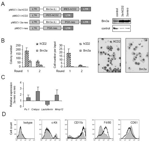
Figure 4. High Brn3a expression promotes myeloid differentiation.

A, left panel) Retroviral plasmids encoding Brn3a(L) cDNAs under the control of MSCV promoter and co-expressing tailless human CD2 (hCD2) or neomycin resistance marker genes were generated as described in Materials and Methods. A, right panel) Brn3a overexpression from retroviruses in comparison with pMSCV-neo (control) was confirmed by immunoblotting of packaging cells. B) Semi-solid colony-forming assays of murine haematopoietic progenitor cells from day E13.5 foetal liver transduced with pMSCV-hCD2 (hCD2) or pMSCV-3a-hCD2 (Brn3a-hCD2) viruses, 1 × 10⁴ FACS-sorted hCD2+ cells plated into each culture 2 days after infection. Colony number per plate and cell number were monitored at rounds 1 and 2, cell morphology (× 40 magnification) at round 1. C) Real-time PCR analysis of gene expression as indicated relative to that of 18s in progenitor cells transduced with pMSCV-neo (control) or pMSCV-3a-neo (Brn3a) and cultured for 6 days under G418 selection. D) Cell-surface FACS analysis of cells transduced with pMSCV-neo (grey filled histogram) or pMSCV-3a-neo (black line histogram) viruses as in C performed at day 9 post-infection, data are representative of three independent experiments
