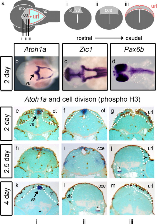
Figure 2.

Atoh1a and proliferation in the zebrafish. Schematic dorsal view (a) of a 5 d embryonic zebrafish shows cerebellar and precerebellar territory in gray, the rhombic lip at the edge of the fourth ventricle in red, and roof plate in light blue. The cerebellar or upper rhombic lip (url) is the portion that borders the cerebellum (cb), which lies between the dorsal lobes of the optic tectum (ot) of the midbrain (mb) and hindbrain (hb). Lines indicate the level of sections through the presumptive valvulus (i) (va), the corpus of the cerebellum (ii) (cce), and the upper rhombic lip (iii). At 2 d (48 h), before the corpus is defined, Atoh1a (blue) is expressed throughout the rhombic lip of both the cerebellum and hindbrain (b) in the same domain as Zic1 (c). Pax6a (data not shown) and Pax6b (d) are not expressed in the rhombic lip. In transverse section, proliferative (phosphohistone H3; brown), Atoh1-positive cells are seen at the valvulus (e), flanked by dividing cells in the dorsal midbrain comprising the optic tectum (ot), at the cerebellar midline (f) and at the Atoh1a-positive upper rhombic lip (g). At 2.5 d, proliferating cells are seen in the Atoh1a-positive presumptive valvulus (h) and in regions in which Atoh1 is downregulated: the midline of the main corpus cerebellar (i) and the url (j). A similar pattern is observed at 4 d with infrequent division at the cerebellar midline (k–m). Dotted white lines indicate the boundaries of cerebellar territory.
