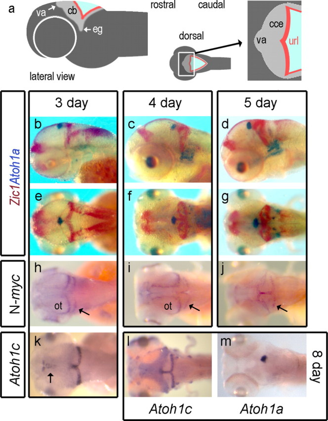
Figure 3.

Atoh1a and Atoh1c in relation to granule cell formation in zebrafish. Schematic lateral view of a zebrafish embryo (a) and expanded dorsal representation of the cerebellum showing the position of the valvulus (va) relative to the corpus of the cerebellum (cce) and upper rhombic lip (url), whose lateral extensions comprise the granule cell eminences (eg). In b–g, Atoh1a (blue) is contrasted with Zic1 (red) in lateral view at 3 d (b), 4 d (c), and 5 d (d) and dorsal view at 3 d (e), 4 d (f), and 5 d (g). N-myc is expressed within caudal margin of the optic tectum (ot) and upper rhombic lip at 3 d (h), expanding into the midline of both at 4 d (i) to include the valvulus at 5 d (j). Atoh1c-positive cells (k) are found in the upper rhombic lip from 3 d onward. At 8 d (l), Atoh1c expression is also seen in anterior lateral line neuromasts and occupies a complementary domain to valvular Atoh1a expression (m) in the cerebellar midline.
