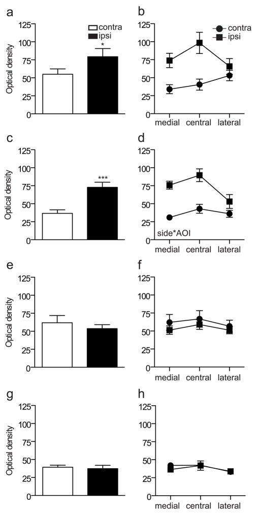
Figure 8.
Quantitation of immunostaining intensity for GFRα3 and CGRP in the L5 dorsal horn following sciatic nerve injury. Optical density measurements in spinal cord sections immunostained for GFRα3 are shown in a–d, whereas data from sections stained for CGRP are shown in e–h. a, Optical density measurements pooled from all superficial areas of interest (AOI 1–3) on each side showed a main effect of CCI (*P = 0.013, F1, 5 = 14.031), with an increase in GFRα3-IR ipsilateral to injury. b, Optical density measurements from each AOI in the superficial dorsal horn did not detect a significant interaction between side and AOI after CCI. c, Optical density measurements pooled from all superficial AOI (1–3) on each side showed a main effect of transection (***P <0.0001, F1, 5 = 85.214), with an increase in GFRα3-IR ipsilateral to injury. d, Optical density measurements from each AOI in the superficial dorsal horn detected a significant interaction between side and AOI (P = 0.014, F1.359, 6.793 = 9.697). e, There was no effect of CCI on optical density measurements of CGRP-IR when all AOIs were pooled and no interaction was detected between side and AOI (f). g, There was no effect of transection on optical density measurements of CGRP-IR when all AOIs were pooled and no interaction was detected between side and AOI (h). Data represent mean ± SEM (n= 6 rats per group), analysed by ANOVA. CGRP, calcitonin gene-related peptide; GFR, GDNF family receptor; IR, immunoreactivity.

