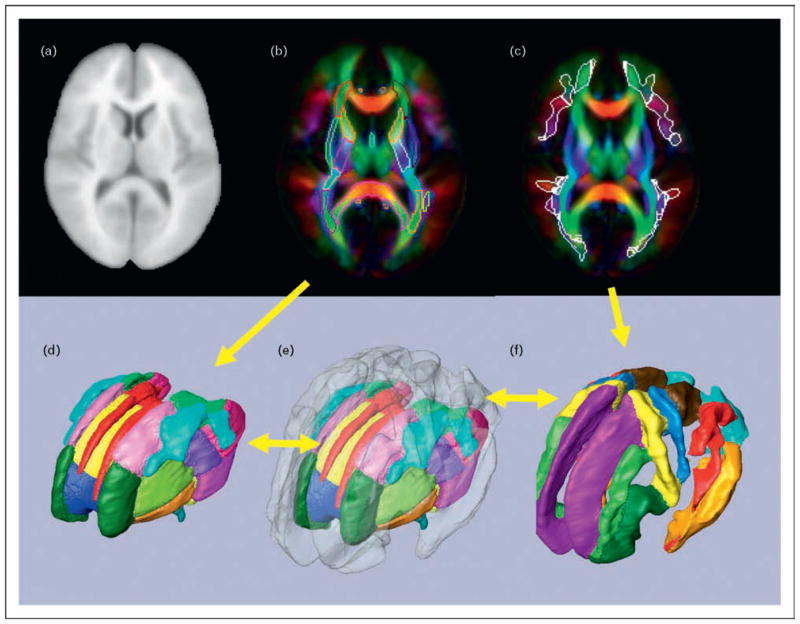
Figure 5. An example of a population-based atlas and parcellation of white matter structures that are reproducible among normal participants.
(a) ICBM-152 atlas created from the T1-weighted images of 152 normal participants. (b) ICBM-DTI-81 atlas created from the diffusion tensor imaging (DTI) of 81 normal participants. The core white matter structures are delineated manually. (c) Superficial white matter regions defined by probability (white lines). (d) Three-dimensional presentation of hand-segmented, core white matter structures. (e) Three-dimensional presentation of the entire white matter structure defined by 60% white matter probability. (f) Three-dimensional presentation of the superficial white matter structures.

