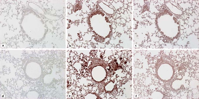
Fig. 11.
Immunohistochemical analysis of SR-A and MARCO expression in whole-lung sections from TLR9+/+ and TLR9–/– mice on day 28 after conidium challenge. Whole-lung tissue sections from Aspergillus-sensitized and challenged TLR9+/+ and TLR9–/– mice were stained using routine immunohistochemical techniques. Representative whole-lung sections from TLR9+/+ (a–c) and TLR9–/– mice (d–f) mice stained with IgG control (a, d), anti-SR-A antibody (b, e), or anti-MARCO antibody (c, f) are shown. Receptor expression stains brown with this immunohistochemical procedure (original magnification: ×200).

