Figure 4.
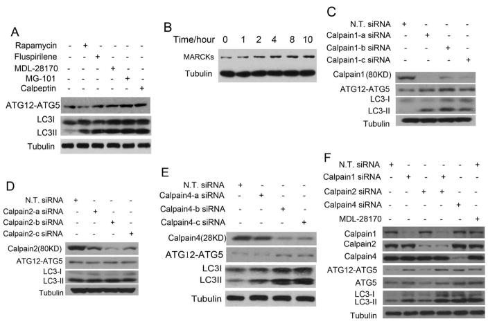
The effects of inhibiting calpains on the levels of ATG12-ATG5 and LC3II in H4 cells. (A) H4 cells were treated with indicated concentrations of rapamycin (0.25 μM), fluspirilene (10 μM), MDL-28170 (10 μM), MG-101 (10 μM) and Calpeptin (10 μM), respectively, for 4 hours. 0.1% DMSO was used as a negative control. The cell lysates were harvested and analyzed by western blotting using anti-ATG12 and anti-LC3. Anti-tubulin was used as a loading control. (B) H4 cells were treated with 10μM fluspirilene for indicated length of time. The cell lysates were harvested and analyzed by western blotting with anti-MARCKs antibodies. Anti-tubulin was used as a loading control. (C–E) H4 cells were transfected with indicated siRNAs for 72 hrs and no targets siRNA (N. T. siRNA) was used as negative control. The cell lysates were harvested and analyzed by western blotting using anti-calpain1 (C), anti-calpain2 (D), anti-calpain4 (E), anti-ATG12 and anti-LC3 (C–E). Anti-tubulin was used as loading controls. (F) H4 cells were transfected with siRNAs indicated for 72 hrs or treated with MDL-28170 (10 μM) for 4 hrs and the cell lysates were harvested and analyzed by western blotting using anti-Calpain1, anti-Calpain2, anti-Calpain4, anti-ATG12, anti-ATG5 and anti-LC3 antibody. Anti-tubulin was used as a loading control.

