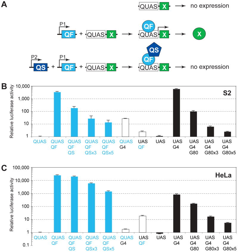
Figure 1. Characterization of the Q-system in Drosophila and Mammalian Cells.
(A) Schematic of the Q repressible binary expression system. In the absence of the transcription factor, QF, the QF-responsive transgene, QUAS-X, does not express X (top). When QF and QUAS-X transgenes are present in the same cell where QF is expressed (promoter P1 is active), QF binds to QUAS and activates expression of gene X (middle). When QS, QF and QUAS-X transgenes are present in the same cell, and both P1 and P2 promoters are active, QS represses QF and X is not expressed (bottom).
(B) Characterization of the Q-system in transiently transfected Drosophila S2 cells. Relative luciferase activity (normalized as described in Extended Experimental Procedures) is plotted on a logarithmic scale on the y-axis, with QUAS-luc2 alone set to 1. Error bars are SEM. Plasmids used for transfections are noted below the x-axis. QUAS, pQUAS-luc2 reporter; QF, pAC-QF; QS, pAC-QS; UAS, pUAS-luc2 reporter; G4, pAC-GAL4; G80, pAC-GAL80; ×3 and ×5, 3 and 5-fold molar excess of QS over QF or GAL80 over GAL4.
(C) Characterization of the Q-system in transiently transfected human HeLa cells. Explanations and abbreviations as in (B) except: QF, pCMV-QF; QS, pCMV-QS; G4, pCMV-GAL4; G80, pCMV-GAL80.
Figure S1 shows the effects of quinic acid on the Q and GAL4 systems.

