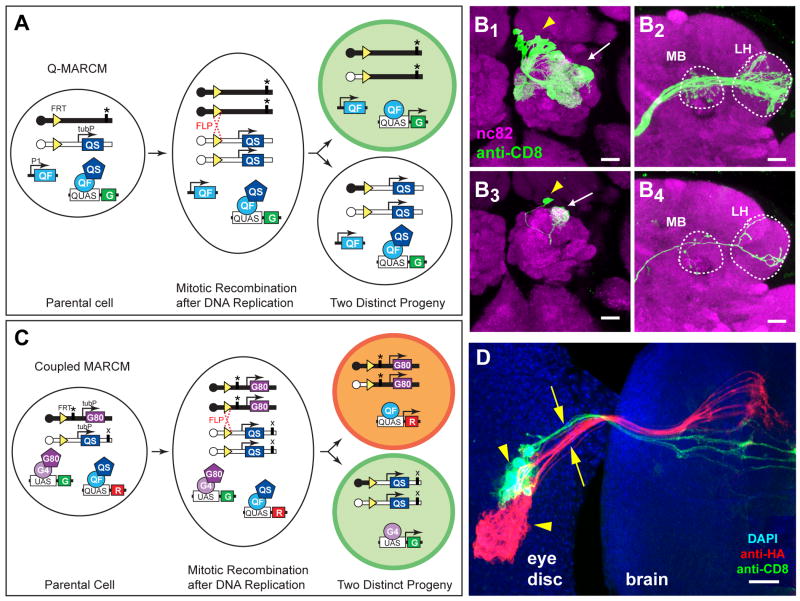
Figure 3. Q-MARCM and Coupled MARCM.
(A) Scheme for Q-MARCM. FLP/FRT mediated mitotic recombination in G2 phase of the cell cycle (dotted red cross) followed by chromosome segregation as shown causes the top progeny to lose both copies of tubP-QS, and thus becomes capable of expressing the GFP marker (G) activated by QF. It also becomes homozygous for the mutation (*). QF and QUAS reporter transgenes can be located on any other chromosome arm. P1, promoter 1. tubP, tubulin promoter. Centromeres are represented as circles on chromosome arms.
(B) Q-MARCM clones of olfactory PNs visualized by GH146-QF driven QUAS-mCD8-GFP. (B1-B2) Confocal images of an anterodorsal neuroblast clone showing cell bodies of PNs (arrowhead), their dendritic projections in the antennal lobe (arrows) and axonal projections in the MB and LH (outlined). (B3-B4) Confocal images of a single cell clone showing the cell body of a DL1 PN (arrowhead), its dendritic projection into the DL1 glomerulus (arrow) of the antennal lobe and its axonal projection in the MB and LH (outlined).
(C) Scheme for coupled MARCM. The tubP-GAL80 and tubP-QS transgenes are distal to the same FRT on homologous chromosomes. Mitotic recombination followed by specific chromosome segregation produces two distinct progeny devoid of QS or GAL80 transgenes, respectively, and therefore capable of expressing red (R) or green (G) fluorescent proteins, respectively. QF and GAL4 transgenes (not diagramed), as well as QUAS and UAS transgenes, can be located on any other chromosome arm. ‘*’ and ‘x’ designate two independent mutations that can be rendered homozygous in sister progeny.
(D) A coupled MARCM clone of photoreceptors, showing clusters of cell bodies (arrowheads) in the eye imaginal disc and their axonal projections (arrows) to the brain. The green clone was labeled by tubP-GAL4 driven UAS-mCD8-GFP; the red clone was labeled by ET40-QF driven QUAS-mtdT-HA. Blue, DAPI staining for nuclei. Image is a z-projection of a confocal stack.
Scale bars: 20 μm.
Figure S3 shows the lack of cross-activation and cross-repression of the Q and GAL4 systems in vivo, and a schematic of independent double MARCM.

