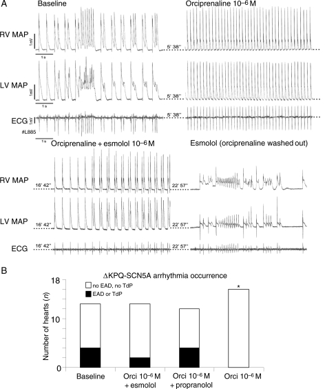
Figure 5.
Antiarrhythmic effect of β-adrenoceptor stimulation in isolated beating hearts. (A) Representative recording of two simultaneous monophasic action potentials (MAP) and ECG during afterdepolarizations and short torsades de pointes (TdP) episodes at baseline after AV block (top left). TdP are suppressed by β-adrenoceptor stimulation (orciprenaline, top right). Ectopy reappears during co-infusion of orciprenaline and β-adrenoceptor-blocker esmolol, and pause-triggered TdP occurs during infusion of esmolol alone. (B) Number of ΔKPQ-SCN5A hearts with arrhythmias at baseline, with β-adrenoceptor-blockers, during co-infusion of β-adrenoceptor-blockers and orciprenaline, and with orciprenaline alone; *P < 0.05 vs. baseline (number of hearts depicted as columns).

