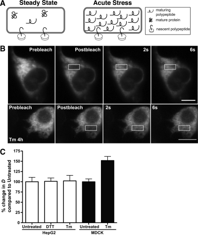
Figure 1.
Misfolded protein stress and the crowdedness (viscosity) of the ER lumen. (A) Proposed model of ER lumen crowdedness during steady state (left) and acute misfolded protein stress (right). At steady state, the lumen contains few immature polypeptides. Acute protein misfolding could fill the lumen with incompletely folded proteins, which could obstruct free diffusion of luminal ER proteins. (B) FRAP of HepG2 cell expressing ER-RFP, untreated or treated with 5 μg/ml Tm. Scale bar, 10 μm. (C) FRAP D values for HepG2 cells or MDCK cells transiently expressing ER-GFP or ER-RFP, untreated or treated with 5 mM DTT for 1 h or 5 μg/ml Tm for 4 h. The mean D values of the two treatments were compared with untreated cells, were converted to a percentage relative to untreated cells for ease of comparison, and are not significantly different for HepG2 cells, but are significant (p = 0.0002) for MDCK cells. D values are provided in Table 1.

