Figure 2.
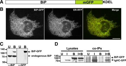
Construction and Characterization of BiP-GFP. (A) Illustration of fusion of hamster BiP in-frame to monomeric GFP followed by a KDEL ER retrieval motif. (B) BiP-GFP colocalizes (yellow in merge panel) with ER-RFP in the ER of a cotransfected Cos7 cell. (C) BiP-GFP (top band) migrates slower than endogenous BiP (bottom band in both untransfected (lane U) and transfected (lane B) cells in an immunoblot of transiently transfected MDCK cell lysate (B lanes) stained with anti-BiP or anti-GFP. (D) BiP-GFP associates with IgHC fused to GFP and is pulled down when coexpressed in cells with IgG heavy chain. Results are displayed in an immunoblot probed with anti-GFP. In the left lanes (1–4), transiently transfected Cos7 cell lysates contain no band (untransfected, (U), IgHC-GFP (I), BiP-GFP (B) or both (I+B). Only I and I+B contain bands of IgHC-GFP, with BiP-GFP in the I+B lane, too. No material is present in U or B pulldown lanes, demonstrating the specificity of the interaction. Molecular-weight-marker positions are indicated to left of blots.

