Figure 8.
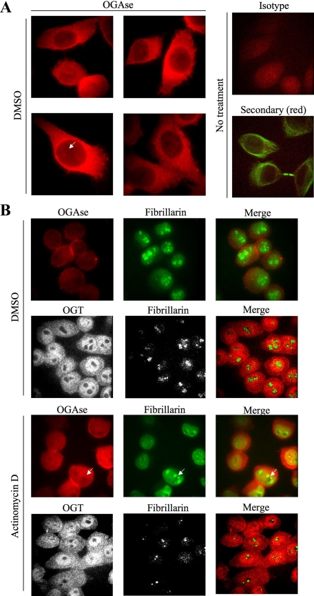
Nucleolar stress disrupts staining of OGAse in the nucleus of cells. (A) HeLa cells were grown on coverslips, fixed, permeabilized, and stained for immunofluorescence confocal microscopy with OGAse (red) antibody. Nuclear OGAse staining shows a gradient from nucleoplasm to nucleoli (white arrow). Isotype (red) and secondary (red, tubulin shown in green) controls are included. Magnification, ×100. All pictures were exposed for equal times (50 ms) and subjected to the same brightness/contrast adjustments. (B) HeLa cells were subjected to treatments as indicated, processed as in A, and double-stained with OGAse (red) and fibrillarin (green) or OGT (red) and fibrillarin (green) antibodies. OGAse staining in the nucleus follows the same pattern as disrupted fibrillarin (white arrows). Magnifications, ×63 (OGT) and ×100 (OGAse). All pictures in each panel were exposed for equal times and subjected to the same brightness/contrast adjustments. Data shown are representative of at least three independent experiments.

