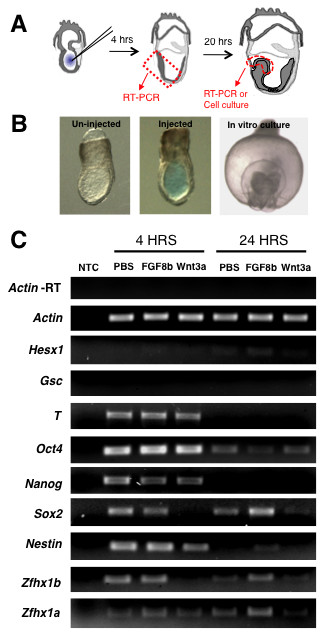
Figure 1.

Microinjection of FGF or Wnt3a modulates neural gene expression in the ectoderm and neural plate. (A) Schematic of experimental setup. (B) Embryos at 7.0 dpc that were either uninjected (left) or injected with a PBS solution containing 0.05% Fast Green dye (middle) within the pro-amniotic cavity. On the far right is an anterior view (dorsal to the top) of an injected embryo after 24-hour whole embryo culture showing normal progression through gastrulation. (C) Semi-quantitative RT-PCR of tissue isolated at 4 hours or 24 hours post-injection and assayed for expression of neurectoderm specific genes (Nestin, Sox2, Hesx1, Zfhx1b, Zfhx1a), pluripontentiality genes (Oct4 and Nanog), or mesodermal genes (Brachyury (T), Goosecoid (Gsc)). Actin gene expression was used for relative quantification and Actin-RT was a control for genomic DNA contamination. Experiment was performed with two repeats on three biological samples, five embryos per sample.
