Figure 6.

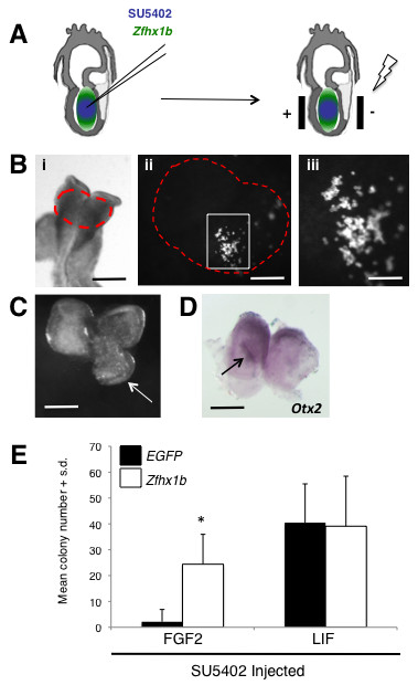
Zfhx1b is sufficient to induce a definitive neural stem cell identity in the anterior ectoderm. (A) Schematic of the microinjection and electroporation procedures. (B) Electroporated embryo after 24-hour culture; the red dashed area in (i) denotes where the EGFP reporter can be visualized in cells of the anterior neural plate (scale bar: 250 μm). (ii) Magnified area of the dorsal head from (i) (scale bar: 50 μm); white box denotes region of transfected cells. (iii) Magnified area in the white box from (ii) (scale bar: 25 μm). (C) Over-expression of Zfhx1b is sufficient to induce ectopic neural plate/ridge-like tissues (arrow) in 75% of the embryos (n = 8; scale bar: 200 μm). (D) Ectopic neural plate/ridge-like tissue (arrow) expresses high levels of the neural marker Otx2 (scale bar: 200 μm). For (C,D), dorsal view of the head region, anterior to the top. (E) Number of NSC colonies derived from 8.5-dpc anterior neural plate cells over-expressing in vivo the control construct alone (EGFP) or EGFP + Zfhx1b (four separate experiments; n = 3 embryos (expressing control reporter construct) and n = 5 embryos (expressing EGFP + Zfhx1b construct); two replicates per culture condition per treatment). The asterisk denotes a statistically significant result (P < 0.05) compared to the transfection control. S.d., standard deviation.
