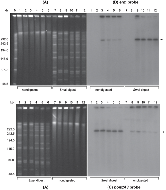
Figure 3. Confirmation of plasmid pBotCDC-A3-Erm transfer from C. botulinum strain CDC-A3580s1 to strain LNT01 by PFGE and Southern hybridization analysis.
(A) Ethidium bromide stained PFGE of C. botulinum DNA samples: SmaI digested DNA of C. botulinum strain LNT01 (Lanes 1 and 7), CDC-A3 wild type (Lanes 2 and 8), CDC-A3580s1 (Lanes 3 and 9), and LNT01 transconjugants (pBotCDC-A3-Erm) (Lanes 4–6 and 10–12); Lanes 1–6, nondigested DNA samples; Lanes 7–12, SmaI digested DNA samples. Lambda PFG Marker (Lane M), New England Biolabs. The position of the pBotCDC-A3 plasmid is indicated with an arrow. Southern hybridization with: (B) the ermB probe, and (C) the bont/A3 probe. PFGE conditions: 6V/cm, 12°C, 1–26 s pulse time, 24 h.

