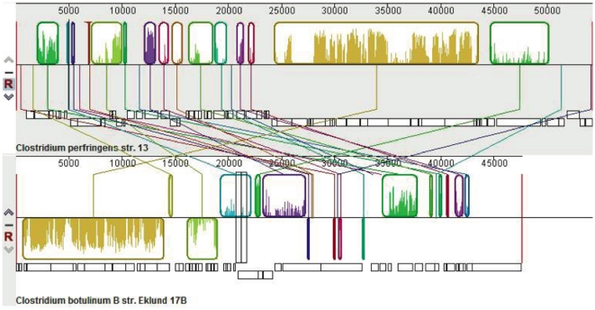
Figure 7. Plasmid alignment of pCLL (C. botulinum strain Eklund 17B) and pCP13 (C. perfringens strain 13).
The alignment has two panels, one for each complete plasmid: pCP13 [top position] and pCLL [bottom position]. The top portions of the panels are composed of colored segments corresponding to the boundaries of locally collinear blocks (LCBs) with lines connecting the homologous blocks in each plasmid. LCBs below a plasmid's centerline are in the reverse complement orientation relative to the reference plasmid (pCP13). The lower portion of the panels represent the predicted open reading frames (ORFs) for the corresponding segments of double stranded DNA with ORFs on top representing top strand and below (bottom strand).

