
Fig. 1.
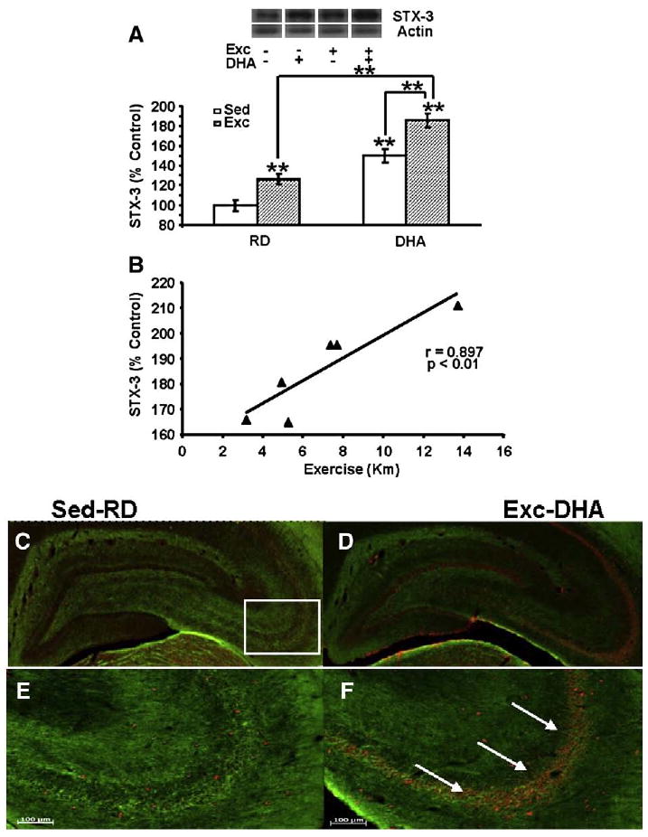
Exercise enhanced the effects of DHA dietary supplementation on STX-3, a protein implicated with the action of DHA on synaptic membrane (A). The values were converted to percent of RD-Sed controls (each value represents the mean latency±SEM, two-way ANOVA, **P<0.01). (B) The levels of STX-3 changed proportionally to the amount of exercise in animals receiving DHA diet (r=0.897, p<0.01). (C–F) Immunofluorescence for STX-3 in coronal sections of the hippocampus shows STX-3 (red, Cy3 secondary antibody) in RD-Sed controls (C, E) and animals receiving combined exercise and diet treatment (DHA-Exc; D, F). (D) and (F) are high magnification photomicrographs (of D and F). A marked increase in STX-3 immunofluorescence was shown in the DHA-Exc group (white arrows, F). Myelinated axons were labeled using immunofluorescence for myelin-associated glycoprotein (green, FITC secondary antibody). RD-Sed: regular diet-sedentary; RD-Exc: regular diet-exercise; DHA-Sed: DHA diet-sedentary; DHA-Exc: DHA diet-exercise.