
Figure 4.
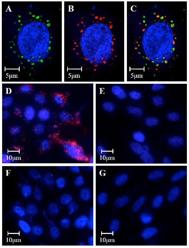
Scanning fluorescence microscopy images demonstrated the co-delivery of (A) the lipid shell to which the hAb conjugated (visualized with green Alexa Fluor 488 dyes) and (B) the PLGA polymeric core (visualized with red Alexa Fluor 647 dyes) to CEA-positive BxPC-3 cells (nucleus stained in blue with DAPI). (C) The overlay of the green and red fluorescence confirmed the integrity of the lipid-polymer hybrid NP structure after being internalized by the target cells. Targeting specificity was confirmed by incubating (D) CEA-positive BxPC-3 cells with anti-CEA hAb targeted NPs, (E) CEA-negative XPA-3 cells with anti-CEA hAb targeted NPs, (F) CEA-positive BxPC-3 cells with non-targeted NPs, and (G) CEA-negative XPA-3 cells with non-targeted NPs.