
Fig. 3. Applying continuous tension with a stage-based force clamp.
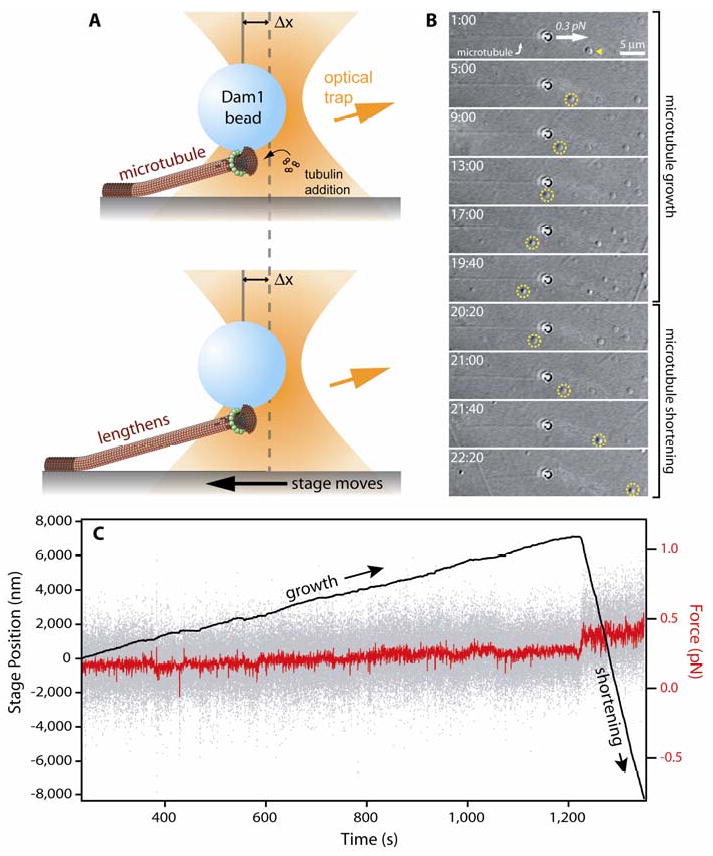
(A) Cartoon showing force clamp operation. A microbead coated with kinetochore proteins (here, the Dam1 complex) is bound to the growing tip of a microtubule. As the microtubule grows and shortens, the stage is moved automatically to keep a fixed offset, Δx, between the trapping laser and the bead, thereby maintaining a constant tensile load on the bead-microtubule interface. Dam1 complexes are depicted here to assemble into a bead-bound ring that encircles the microtubule (35, 36). However, rings are not required for coupling (e.g., see 48, 50) and they are not known to exist in vivo. Thus, the functional importance of rings remains uncertain. (B) Time-lapse images showing a Dam1 complex-coated bead tracking with microtubule growth (1 min:00 sec to 19:40) and shortening (20:20 to 22:20) under constant force (0.25 pN and 0.4 pN, during growth and shortening, respectively). The stage is moved leftward during growth, as evident by a coverslip-adsorbed fiducial (yellow arrowhead, circles), and rightward during shortening. (C) Record of stage position (black trace) and force (instantaneous, gray trace; average, red trace) versus time for the event shown in (B).