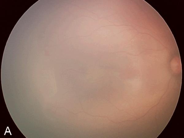
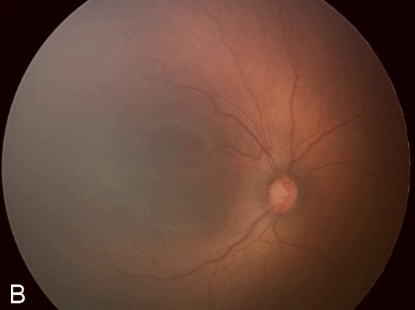
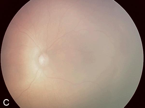
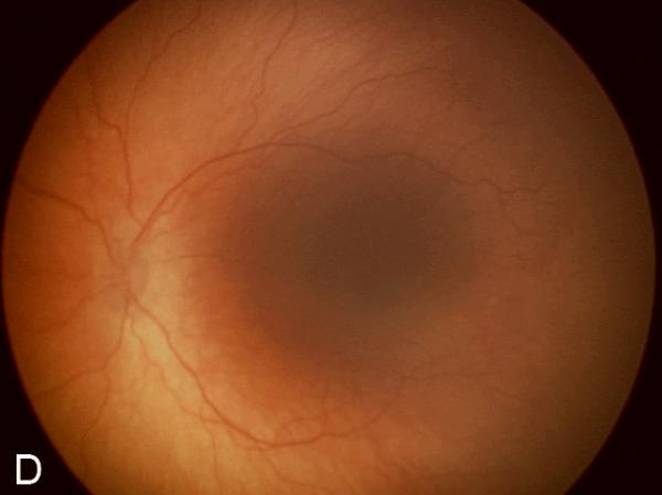
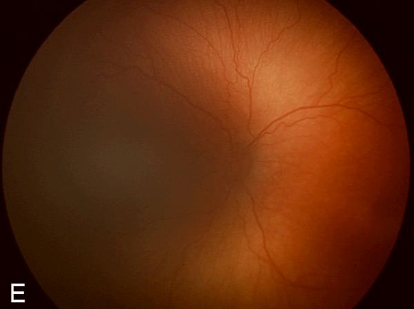
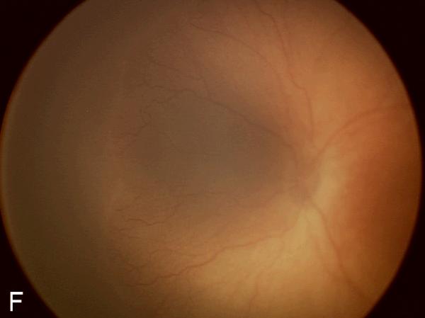
Figure 2. Examples of study images that were frequently misdiagnosed by retinal fellows.
(A), (B), and (C) display temporal, posterior, and nasal images from an infant diagnosed as mild ROP by reference standard exam diagnosis and was diagnosed as type-2 ROP by 5/7 (71%) fellows, and as treatment-requiring ROP by 2/7 (29%) fellows. (D), (E), and (F) display temporal, posterior, and nasal images from an infant diagnosed as type-2 ROP by reference standard exam diagnosis and was diagnosed as no ROP by 2/7 (29%) fellows, as type-2 ROP by 1/7 (14%) fellows, and as treatment-requiring ROP by 4/7 (57%) fellows.

