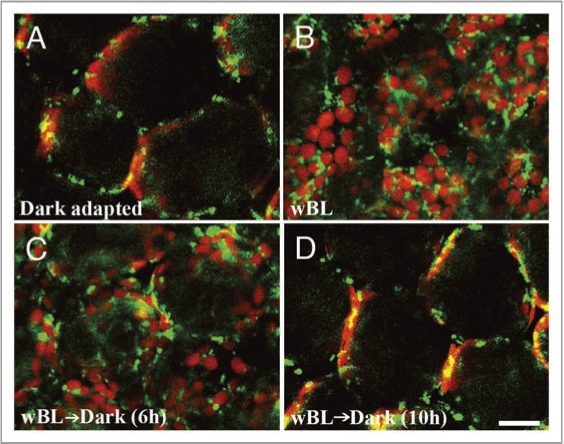
Figure 1.
Distribution of mitochondria and chloroplasts on the outer periclinal regions of palisade mesophyll cells of A. thaliana under different light conditions. Mitochondria (green; GFP) and chloroplasts (red; chlorophyll autofluorescence) were visualized with confocal microscopy after dark adaptation (A), immediately after wBL (470 nm, 4 µmol m−2s−1) illumination for 4 h (B), after dark treatment for 6 h (C) and 10 h (D) following the 4-h wBL illumination, respectively. Bar = 50 µm.

