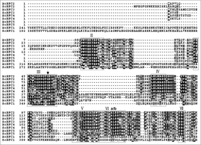
Figure 1.
RFC boxes II to VII of RFC proteins from Arabidopsis thaliana and Saccharomyces cerevisiae. Alignment was carried out using ebi ClustalW (www.ebi.ac.uk/clustalw/). The amino acids enclosed in the red frame indicate RFC boxes II to VII, which are amino acid sequence motifs conserved in all RFC subunits. Box VIa is conserved in the large RFC subunits, and box VIb is conserved in the other proteins. The arrow points to the mutation site of AtRFC3 in rfc3-1 mutant.

