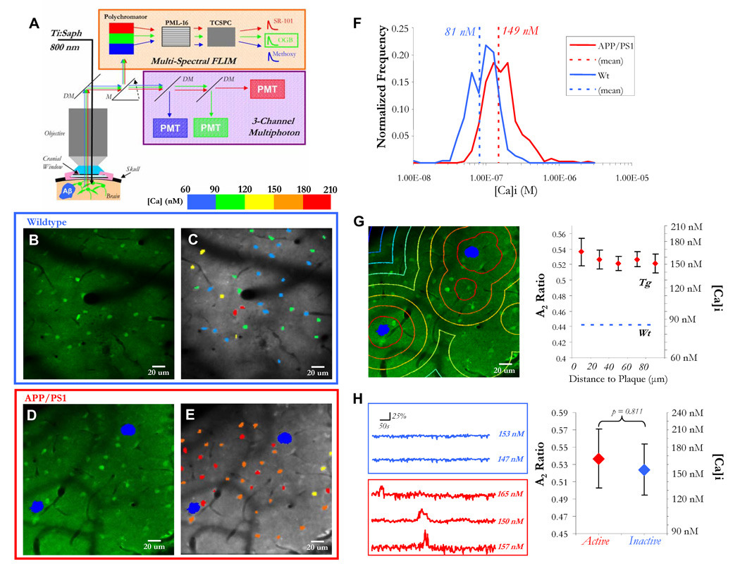
Figure 1. Resting calcium is globally elevated in astrocytic networks.
A) Multiphoton laser illumination simultaneously excited methoxy-XO4 (blue, amyloid-β), OGB (green, neurons and astrocytes), and SR-101 (red, astrocytes) through a cranial window. The resulting fluorescence emission was sent to either (1) a three-channel intensity-based PMT module or (2) a 16-channel multi-spectral FLIM detector. A single-photon counter (tcSPC) recorded fluorescence lifetime data . B–E) Fluorescence decay curves were fit with a calcium bound lifetime (2359 ps) and an unbound calcium lifetime (569 ps) for each pixel. The pixel data were averaged to obtain single- cell calcium levels, depicted with a calibrated colorbar (C,E). Astrocytes in APP/PS1 mice with cortical plaques (in blue, D–E), exhibited significantly higher levels of [Ca]i than in wildtype mice (B–C,F: p < 0.05, Student’s t-test, n = 241 cells in 3 mice (wt), n = 364 cells in 3 mice (Tg)). G) Astrocyte resting [Ca]i did not depend on proximity to a plaque (p = 0.9194, Kruskal-Wallis test, n > 25 cells for each distance group). H) There was no difference in resting calcium between cells that were active versus inactive (p = 0.811, Student’s t-test, n = 209 cells in 3 mice).

