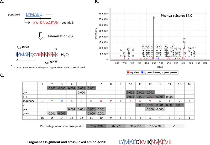
Figure 3. Example of an identification based on only one permutation in the cytochrome P450 2E1/cytochrome b5 complex.
A) Aspartic acid D6 of peptide α is cross-linked to lysine K4 of peptide β. The linearization αβ depicted here covers 80% of the fragment ions. B) and C) Spectrum matching to the linearization αβ. Almost all fragment ions are assigned to this spectrum with a high z-Score using Phenyx. The extent of fragment assignment allows validation of the position of the cross-linked amino acid. Unlike linearization αβ, βα poorly matches to the spectrum with a low z-Score of 3.56 (data-not shown).

