
Figure 3.
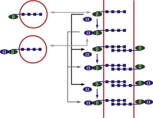
Schematic illustration of the reactions occurring at the cell membrane and leading to Eq. 4. E-cadherin vesicles, either bound to β-catenins alone or to α-catenin-β-catenin complexes, can merge with the membrane or be endocytosed. The two associated light gray arrows correspond to the four on and off rates in Eq. 4. In the presence of cell-cell contact, the E-cadherins on the membrane can bind to E-cadherins on the membrane of the adjacent cell, which is represented by the black arrows for β-catenin-associated complexes, and by the dark gray arrows for α-catenin-β-catenin-associated complexes. These correspond to all the reactions that have rates with an EE superscript in Eq. 4. In addition, α-catenin monomers can bind to E-cadherin-β-catenin complexes present on the membrane, whether or not they are bound to E-cadherins from the adjacent cell. This is represented by the blue vertical arrows and corresponds to the terms in Eq. 4 whose rates have an m,d or m,a superscript. Finally, all protein complexes located on the membrane can be degraded, which is taken into account by the rates labeled with the letter r in Eq. 4 (not represented). We assume a symmetric configuration of the adjacent cell.