
Figure 1.
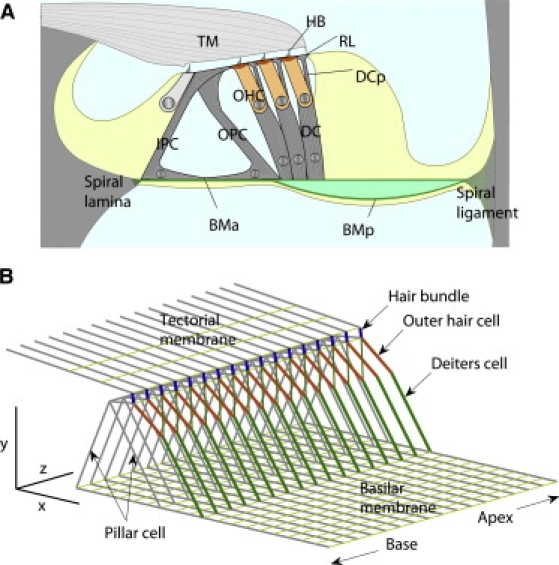
Finite element model of cochlear partition. (A) Radial section of cochlear partition. Organ of Corti sits on basilar membrane (BM), which is divided into arcuate (BMA) and pectinate (BMP) zones. BMA supports inner and outer pillar cells (IPC and OPC) and BMP supports Deiters' cell (DC) and outer hair cell (OHC). Reticular lamina (RL) comprises tops of pillar cells, OHC cuticular plates, and tops of DC phalangeal processes (DCP). (B) Structural components of cochlear partition represented by beam elements allowing elongation and bending. The medial and lateral ends of the basilar membrane were hinged at the spiral lamina and spiral ligament and the tectorial membrane (TM) was fixed. Both elements were composed of beam elements aligned in radial and longitudinal directions. Three rows of OHCs and DCs merged into one with Young's modulus and force generation three-times that of single cell. Deiters' cell phalangeal process (DCP) and OHC form two arms of a Y-shape, surmounting the basal part of the DC. Scale bars 40 μm.