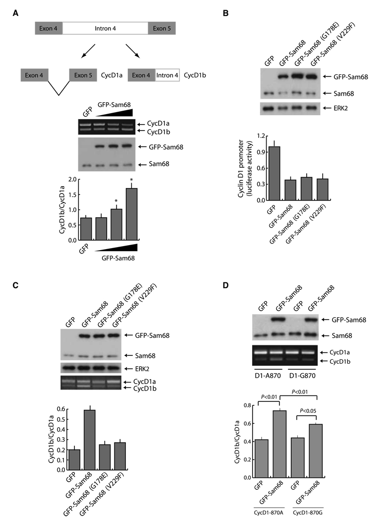
Figure 3.
Sam68 modulates the AS of cyclin D1. A, schematic representation of the cyclin D1 minigene and of the PCR products from the in vivo splicing assay. Top, splicing assay of the cyclin D1 minigene in PC3 cells transfected with the cyclin D1 minigene together with increasing amounts of GFP-Sam68. Cells were harvested 20 h after transfection and processed for RT-PCR and protein extraction. Middle, immunoblot analysis of the same samples of the splicing assay. Bottom, columns, mean of the cyclin D1b/cyclin D1a ratio from three independent experiments; bars, SD. *, P < 0.01. B, cyclin D1 luciferase reporter assay of PC3 cells transfected with pGL3-cyclinD1-luc and GFP-Sam68wt or G178E and V229F mutants. Top, Western blot analysis of the transfected cells; bottom, cell extracts were prepared and assayed for firefly and Renilla luciferase (to normalize for transfection efficiency). Columns, mean of three experiments; bars, SD. C, splicing assay of the cyclin D1 minigene described in A in PC3 cells transfected with the indicated GFP or GFP-Sam68 constructs. Top, immunoblot analysis of the same samples of the splicing assay; bottom, PCR from the splicing assay with the cyclin D1 minigene. Columns, mean of the cyclin D1b/cyclin D1a ratio from three independent experiments; bars, SD. D, in vivo splicing assay of cyclin D1 minigenes 870G or 870A in the presence of GFP-Sam68. PC3 cells were transfected with the indicated constructs and collected 30 h after transfection for RNA and protein extraction. Top, immunoblot analysis of the transfected cells. Middle, PCR analysis for cyclin D1a and cyclin D1b. Bottom, densitometric analysis of three different experiments. Columns, mean; bars, SD. P value is indicated.

