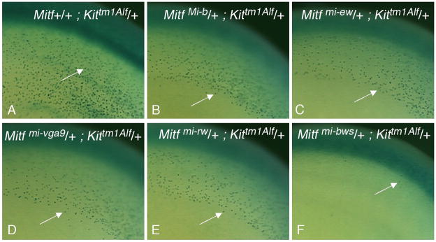
Fig. 3.
Tracking of β-Gal-positive melanoblasts in embryogenesis. Embryos of the indicated genotypes were harvested at 12.5 days of gestation and processed for β-Gal labeling. Arrows point to individual β-Gal-positive melanoblasts in the dorsolateral migration pathway underneath the surface ectoderm in the trunk area. (A-E) Note similar distribution but slightly reduced numbers and densities of β-Gal-positive melanoblasts in embryos carrying a mutant Mitf allele together with the Kittm1Alf allele as opposed to a wild-type Mitf allele along with the Kittm1Alf allele, consistent with earlier findings that Mitf gene dosage affects melanoblast numbers early in development (Hornyak et al., 2001). (F) Melanoblasts in embryos double heterozygous for Mitfmi-bws and Kittm1Alf are very sparse and largely restricted to the area over the neural tube.

