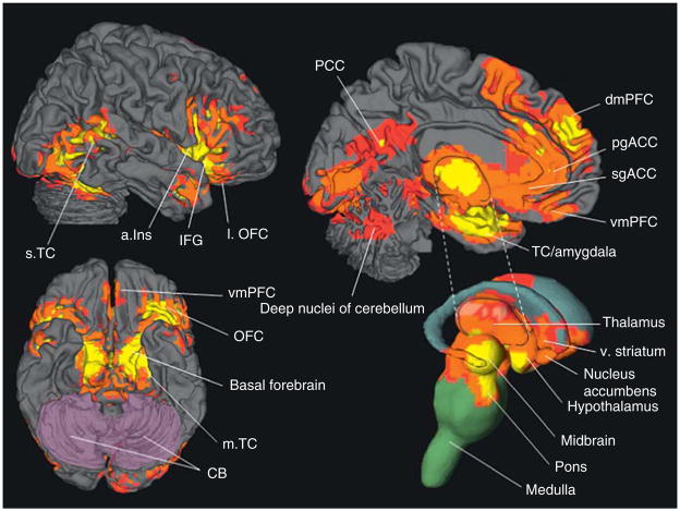
Figure 4.2.
The observed neural reference space for core affect. 165 neuroimaging studies of emotion (58 using PET and 107 using fMRI) published from 1990 to 2005 were summarized in a multilevel meta-analysis to produce the observed neural reference space for emotion (Wager et al., 2008). These areas include (from top left, clockwise) anterior insula (aIns), lateral OFC (lOFC), pregenual cingulate cortex (pgACC), subgenual cingulate cortex (sgACC), ventral medial prefrontal cortex (vmPFC), temporal cortex/amygdala (TC/Amygdala), thalamus, ventral striatrum (v Striatum), nucleus accumbens, hypothalamus, midbrain, pons, medulla, OFC, and basal forebrain. Other areas shown in this figure (e.g., inferior frontal gyrus (IFG), superior temporal cortex (sTC), dorsal medial prefrontal cortex (dmPFC), posterior cingulate cortex (PCC), medial temporal cortex (mTC), and cerebellum (CB)) relate to other psychological processes involved with emotion perception and experience. (See online version of the chapter for color figure).

