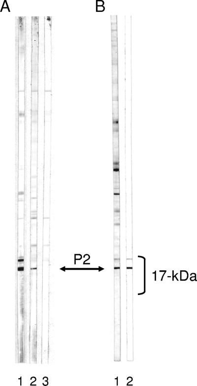
FIG. 4.
Identification of CpP2 band. (A) Total antigen C. parvum PVDF strips were incubated with serum from a mouse that was immunized with purified rCpP2 (lane 1), monoclonal antibody 1D6 raised against rCpP2 (lane 2), or buffer alone (lane 3). Bound IgG antibodies were visualized by use of a biotinylated rat anti-mouse IgG monoclonal antibody and developed as described in the legend to Fig. 1. (B) Total antigen C. parvum PVDF strips were incubated either with serum from a Haitian donor (lane 1) or with human serum antibodies that were eluted from purified rCpP2 (lane 2). Bound IgG antibodies were visualized as described in the legend to Fig. 1. The location of the CpP2 band is indicated by the double-headed arrow.

