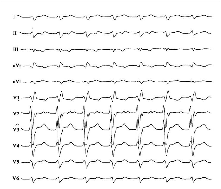
Figure 2.

12-lead-ECG of a patient with wide QRS complex tachycardia. The tracing shows right bundle branch block morphology. Note the typical QRS features in leads V1 and V6 (triphasic appearance of V1, R to S ratio of less than 1 in V6). The AV dissociation and the northwest axis are also helpful clues for the diagnosis of ventricular tachycardia
