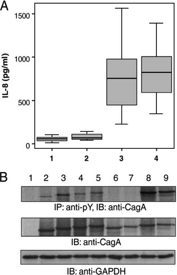
FIG. 1.

(A) Levels of secreted IL-8 following infection of gastric epithelial AGS cells with H. pylori clinical strains (1, cagA negative; 2, cagPAI defective; 3, 1 EPIYA-C repeat; 4, ≥2 EPIYA-C repeats). No difference was observed between cagA-negative and cagPAI-defective strains (U = 32.500 and P = 0.201 by the Mann-Whitney U test). cagA- and cagPAI-positive strains induced higher levels of IL-8 than cagPAI-defective strains (U = 0.000 and P < 0.0001 by the Mann-Whitney U test), irrespective of the number of EPIYA-C sites (U = 224.500 and P = 0.627 by the Mann-Whitney U test). (B) Tyrosine phosphorylation and expression patterns of CagA protein following infection of AGS cells with representative H. pylori clinical strains. CagA tyrosine phosphorylation was detected by immunoblotting (IB) following immunoprecipitation (IP) with PY20 antiphosphotyrosine antibody. The expression of GAPDH (glyceraldehyde-3-phosphate dehydrogenase) was utilized as a total protein loading control. Lanes: 1, CagA-negative clinical isolate; 2 to 5, CagA-positive isolates with functional cagPAI harboring 2 (AB), 3 (ABC), 4 (ABCC), and 5 (ABCCC) motifs in CagA, respectively; 6 and 7, CagA-positive H. pylori strains carrying 3 (ABC) and 4 (ABCC) EPIYA motifs with defective cagPAI, respectively, as depicted by the absence of phosphorylated CagA; 8 and 9, CagA-positive strains with 5 (ABCCC) EPIYA motifs.
