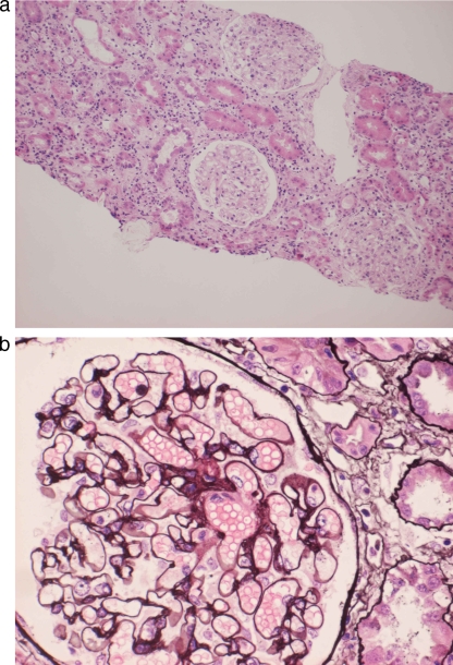
FIG. 3.
(a) Light microscopy finding of interstice showing massive inflammatory cell infiltration (hematoxylin and eosin staining; magnification, ×100). (b) Light microscopy finding of a glomerulus showing glomerular basement membrane (GBM) thickening and small cystic spaces in the GBM (periodic acid-Schiff-methenamine staining; magnification, ×400).

