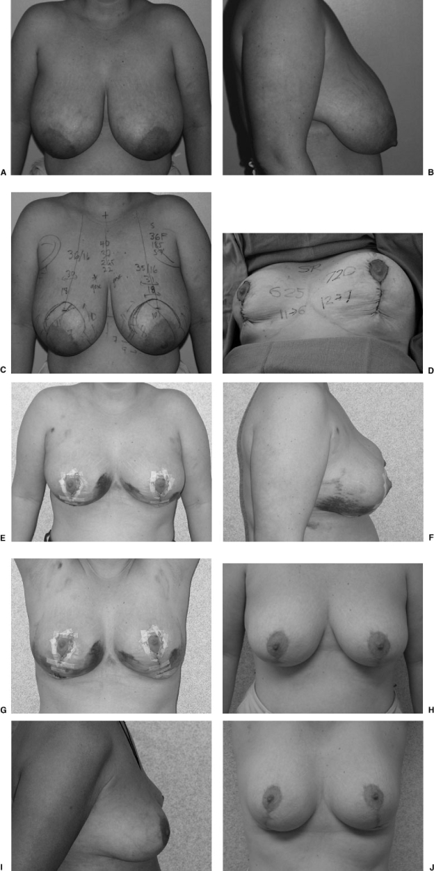
Figure 10.
(A) Preoperative frontal view of moderate-sized breast reduction (34-year-old patient who wore a 36F brassiere, 185 lb, 5′7″). (B) Preoperative lateral view. (C) Preoperative view with markings (many of these measurements are performed for statistical analysis only). The most important markings are noting the level of the inframammary fold (and therefore the new nipple position vertically), the breast and chest wall meridian (and therefore the new nipple position horizontally), as well as the areolar opening, the skin resection pattern, and the medial pedicle design. (D) Intraoperative view at completion of the vertical approach using the medial pedicle. From the right breast, 625 g were removed; 720 g were removed from the left breast. She also had 400 mL of fat removed from the lateral chest wall and preaxillary areas with some contouring of the lower portion of the breasts. Surgery time was 90 minutes. I now gather this incision far less than shown in this photograph. (E) Frontal view at 10 days postoperatively. (F) Lateral view at 10 days postoperatively. (G) Arms up view at 10 days. The results do not necessarily take a long time to settle postoperatively. (H) Frontal view at 15 months postoperatively. (I) Lateral view at 15 months postoperatively. (J) Arms up view at 15 months postoperatively.

