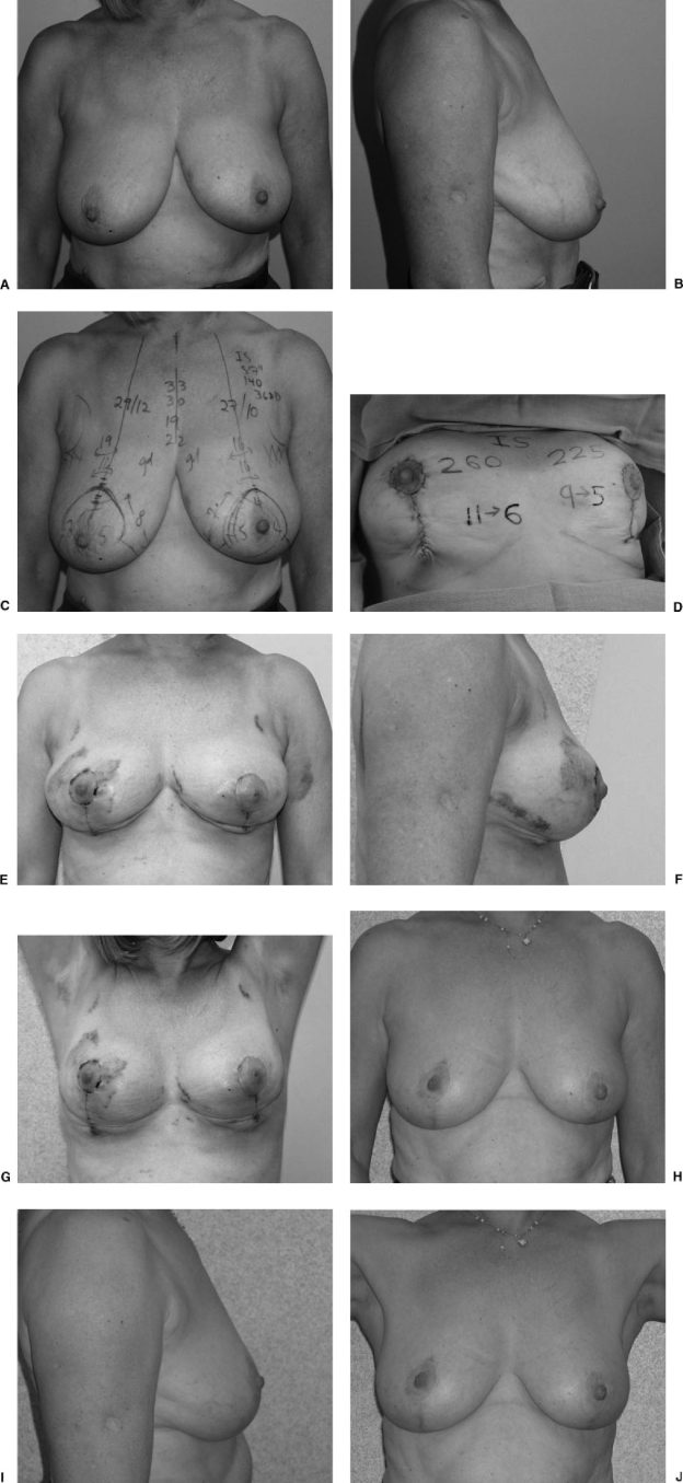
Figure 11.
(A) Preoperative frontal view of small breast reduction (56-year-old patient who wore a 36DD brassiere, 140 lb, 5′7″). (B) Preoperative lateral view. (C) Preoperative view with markings. (D) Intraoperative view at completion of the vertical approach using the medial pedicle. From the right breast, 260 g were removed; 225 g were removed from the left breast. An additional 200 mL of fat was removed from the lateral chest wall and preaxillary areas with some contouring of the lower portion of the breasts. Surgery time was 90 minutes. I now gather this incision far less than shown in this photograph. (E) Frontal view at 2 weeks postoperatively. (F) Lateral view at 2 weeks postoperatively. (G) Arms up view at 2 weeks. The results show some puckering and irregularities. A preoperatively informed patient accepts this and realizes that some time may be needed before it settles. (H) Frontal view at 2 years postoperatively. Note that the nipple is higher on the larger side; it should have been designed at a lower position. (I) Lateral view at 2 years postoperatively. (J) Arms up view at 2 years postoperatively.

