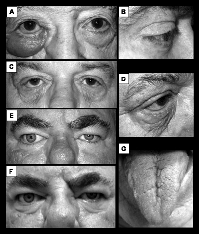
Figure 2.
(A) Unilateral eyelid swelling with erythematous and translucent skin as well as conjunctival swelling (chemosis). (B) Eyelash ptosis of floppy eyelid syndrome with subtle preseptal eyelid edema in both upper and lower eyelids. (C, D) Atopic dermatitis with eyelid skin erythema, thickening, and lost elasticity leading to eyelid retraction. Note the characteristic vertically oriented rhytids. (E) Rosacea-associated posterior lid margin disease blepharitis with mild chemosis and attendant eyelid edema. Flash photography demonstrates a light reflex from chemosis at the eyelid margin in the left eye. (F) Nonflash photograph of same patient in (E) showing shadowing from the lower eyelid edema. (G) Deeply fissured tongue seen in Melkersson Rosenthal syndrome.

