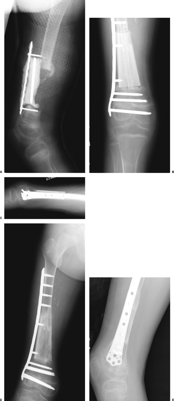
Figure 2.
A case example of a 6-year-old boy who presented after Ewing's tumor resection and reconstruction at an outside institution using femoral allograft. The allograft reconstruction became infected and was replaced with an antibiotic spacer. (A) A lateral radiograph of leg after failed initial treatment. The antibiotic spacer has migrated due to hardware failure. (B) The spacer is removed and reconstructed by Capanna technique with the construct shown in Fig. 1. Fixation is achieved with the use of a laterally placed plate. (D, E) Excellent incorporation of both the fibula and allograft are seen 4 months postoperatively.

