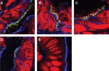
Fig. 5.
Immunofluorescence staining of CR-infected cryosectioned mouse colons. Staining was performed for CR (A, green) and Tir (B–E, green). Filamentous actin was visualized by phalloidin staining (blue), and bacteria and cell nuclei were counterstained with propidium iodide (red). Intimately adhering bacteria with translocated Tir underneath were observed on mouse colons infected with wt CR (B), DBS255(pICC438) (C) and DBS255(pICC327) (D). No adherent bacteria were detected on mouse colons infected with DBS255(pICC55) (E).

