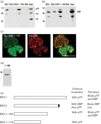
Fig. 3.
(a, b) Western blot for (a) pTP and (b) DBP following depletion of Ad2-infected cell lysates by B23.1, B23.2, B23 1–160 and BSA. (c) Myc–B23 1–160 and pTP in infected cells. Image was taken by using a Leica confocal microscope with a ×63 oil immersion lens and is of a single focal plane approximately 0.3 μm deep; bar, 10 μm. (d) Purified B23 1–160 was analysed by SDS-PAGE and Coomassie stain for quality. (e) Schematic diagram illustrating the binding and co-localization properties of B23.1, B23.2, B23 1–160 and B23 1–116 in infected cells. Note that the last 2 aa of B23.2 are not equivalent to sequences in B23.1. In the case of B23.1 and B23.2, the co-localization is confirmed by EGFP- and Myc-tagged fusion proteins. For B23 1–160, the localization was shown by Myc-tagged fusion protein only and, for B23 1–116, the co-localization was shown by EGFP-tagged fusion protein only.

