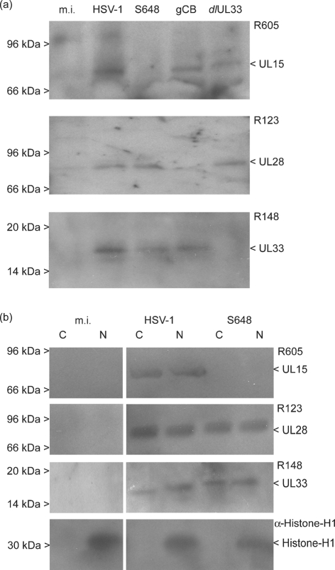
Fig. 3.
Western blot analysis of UL15, UL28 and UL33 expression. (a) Total proteins from mock-infected cells (m.i.) or cells infected with wt HSV-1, S648, gCB or dlUL33 were harvested at 6 h p.i. and the respective proteins detected with antisera against UL15 (R605), UL28 (R123) or UL33 (R148), as indicated. (b) Cytoplasmic (C) and nuclear (N) fractions from m.i. or cells infected with wt HSV-1 or S648 were analysed with the above antisera and an antibody against histone H1. The positions of protein molecular mass markers are shown on the left hand side. It should be noted that the UL15.5 product (30 kDa) is too small to be detected on the blots probed with R605.

