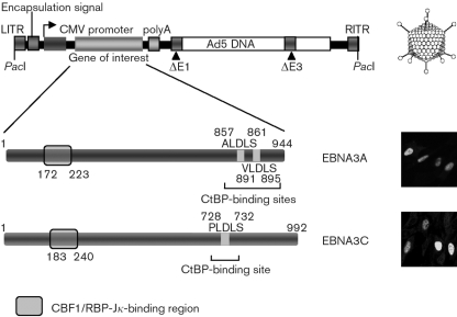
Fig. 1.
Schematic showing the pAdEasy-1 recombinant adenovirus backbone and the EBNA3A and EBNA3C adenoviruses used in this study. The CtBP-binding site(s) (A/V/PLDLS) and predicted CBF1/RBP-Jκ-binding region are shown for EBNA3A and EBNA3C. Immunofluorescence images of infected IMR-90 cells (m.o.i. of 25, 24 h), using antibodies specific for EBNA3A or 3C, are shown in the bottom right panels. CMV, Cytomegalovirus; LITR, left internal repeat; RITR, right internal repeat.

